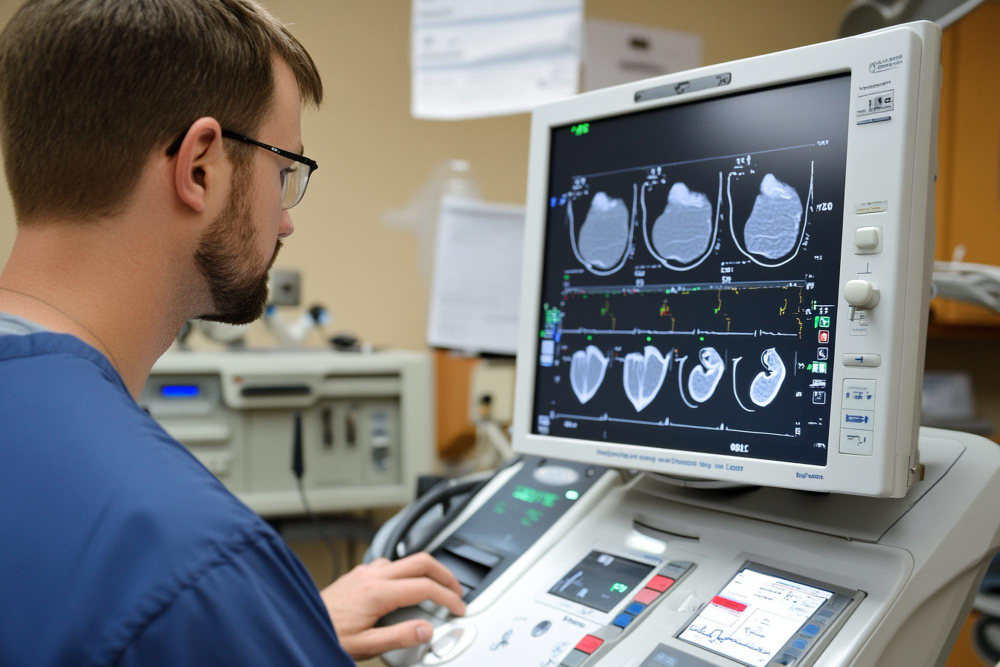
Echocardiography

Echocardiography, also known as an “echo,” is a specialized ultrasound imaging technique that evaluates the structure and function of the heart. It uses high-frequency sound waves to create detailed images, providing crucial insights into cardiac health.

Applications

  • Heart Structure Analysis: Evaluates the size, shape, and movement of heart chambers and walls.
  • Valve Function Assessment: Detects abnormalities in heart valves, such as stenosis or regurgitation.
  • Blood Flow Monitoring: Uses Doppler technology to measure blood flow through the heart and vessels.
  • Disease Diagnosis: Identifies heart conditions such as cardiomyopathy, congenital defects, or pericardial effusion.

Types of Echocardiography

  • Transthoracic Echocardiography (TTE): The standard, non-invasive procedure where a transducer is placed on the chest.
  • Transesophageal Echocardiography (TEE): A specialized test where the transducer is inserted into the esophagus for closer heart imaging.
  • Stress Echocardiography: Performed during or after exercise to assess heart performance under stress.
  • Doppler Echocardiography: Focuses on blood flow and pressure in the heart.

Benefits

  • Non-Invasive: Most echo tests are painless and require no recovery time.
  • Safe: No radiation is involved, making it suitable for repeated use.
  • Real-Time Results: Offers immediate visual insights for diagnosis and treatment planning.